
A portable ultrasound system could make reliable breast imaging more accessible
For people at high risk of developing breast cancer, yearly mammograms may not be enough to detect tumors early. To make earlier diagnosis easier, an MIT team has developed portable detectors based on ultrasound, which could be used much more frequently.
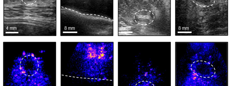
In a new paper, the team reports that they have improved the resolution of the images produced by their system, making it easier to spot potential tumors, as well as cysts and microcalcifications. The researchers also created a user interface that makes it simple to use the ultrasound probe, even for people with no expertise in ultrasonography.
This system, they believe, could not only enable earlier detection, but also allow for long-term monitoring following breast cancer treatment — either in a doctor’s office or at home.
“At each time interval, the computer interface guides you to position the device in exactly the same location, which is important for the longitudinal monitoring of a given tissue. It’s very intuitive and quite easy to use,” says Canan Dagdeviren, an associate professor of media arts and sciences at MIT and the senior author of the study.
Former MIT postdoc Md Osman Goni Nayeem and MIT graduate students Shrihari Viswanath and Hyeokjun Yoon are the lead authors of the paper, which appears today in Nature Communications.
Higher-quality imaging
While many people receive annual mammograms to check for breast cancer, it is possible for cancer to develop in between these annual screenings. These cancers, known as interval cancers, tend to be more aggressive, and they account for 20 to 30 percent of all breast cancer cases.
After losing an aunt to an interval breast cancer in 2015, Dagdeviren was motivated to develop a screening technique that would be more effective on women with dense breast tissue and could be performed more often than mammography. She decided on ultrasound, which uses sound waves to create images of tissue. Ultrasounds are often used to follow up on abnormal mammograms, but current ultrasound technology requires large equipment and a trained operator.
Earlier this year, Dagdeviren’s lab published a study in which they demonstrated a small ultrasound probe attached to an acquisition and processing module that is a little larger than a smartphone. This compact system can create a 3D image of the entire breast by scanning just two or three locations.
In the new Nature Communications study, the researchers reported several advances that allow for higher resolution imaging and greater ease of use.
One key advance is the addition of a “backing layer” to the ultrasound transducer. This layer helps to contain and focus the ultrasound waves, improving the resolution and quality of the resulting images. It also increases the range of soundwave frequencies that can be absorbed, and reduces both acoustical noise and electrical noise, further enhancing the images.
“With the backing layer, the device produces more accurate and sharper images, with a wider operating range of frequencies,” Nayeem says.
To further improve the quality of the images, the researchers designed an algorithm that adaptively performs a process called beamforming. This algorithm allows the system to compensate for differences in the speed at which sound waves travel through different types of tissue, such as skin and fat.
“What we are trying to do is predict the speed of sound properties of the tissue you’re imaging, and then use that to reconstruct the image more accurately. We see up to a 10 percent improvement in the resolution just by applying this technique,” Viswanath says.
The researchers asked 10 volunteers, who were not experts in ultrasound technology, to use the system to try to identify small micro targets embedded in a “tissue phantom” — a gel-like material engineered to mimic human tissue. Participants had a much higher success rate locating the spheres when they used the new system than when they used a traditional ultrasound probe.
A user-friendly system
For the new version of this system, the researchers also created a user interface, displayed on a computer screen, that guides the user to place the probe in the correct location. This could be especially important for tracking progression of treatments such as neoadjuvent therapy, or long-term monitoring of known abnormalities such as fibroadenomas or microcalcifications.
In a trial with seven people, the researchers found that the users were able to accurately place the probe in the correct location each time they did a scan.
“Conventionally, you need an operator to move the probe around the breast, but we made a computer-vision interface for users to do it by themselves. This is very user-friendly and it shows live images on the screen,” Yoon says.
For future versions of this technology, the researchers hope to create an interface that could be used with a cellphone or tablet, making the system easier to carry. In addition to enabling earlier diagnosis, this type of system could make ultrasound more accessible to patients in areas where there aren’t enough trained ultrasound technicians, the researchers say.
Dagdeviren and some of her students now hope to form a company to work toward making the technology commercially available. While breast cancer diagnosis is their first target application, they hope to expand it to many others.
“The technology is so versatile that it can be used for any soft tissue imaging, from ovarian cancer to measuring endometriosis progression, or fetal monitoring,” Dagdeviren says.
The research was funded by the National Science Foundation, the 3M Non-Tenured Faculty Award, the Lyda Hill Foundation, the MIT Media Lab Consortium, and a Tata Center Technology and Design Fellowship.